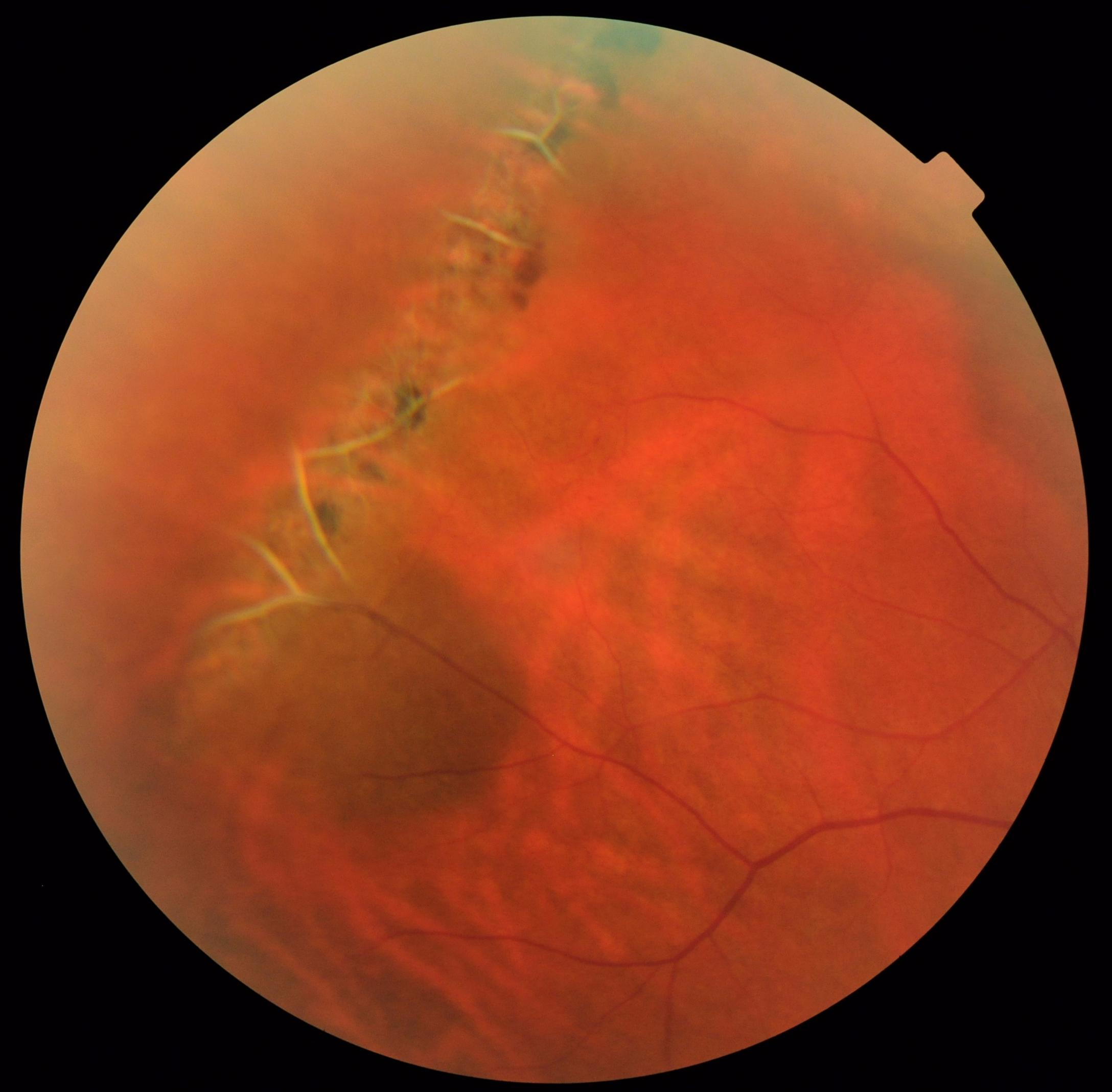

7 Mirror artifacts (inverted artifacts) are often detected in myopic eyes or in the peripheral part of nonmyopic eyes due to the scleral curvature, 6, 7 although they only occur from the Fourier transformation used in OCT systems, including spectral-domain and SSOCT. 6, 7 Of the OCT artifacts, it has been reported that the peripheral part of the OCT image is partially inverted in some cases, 6 which is called as a mirror artifact 6, 7 or an inverted artifact.

It is well known that there are various artifacts in OCT image scans. 4, 5 However, it is still difficult to capture OCT images of the far-peripheral part of the retina. Recently, wide angle swept-source OCT (SSOCT) (Plex Elite 9000, Carl Zeiss Meditec, Dublin, CA) has been able to assess the mid-peripheral retinal structure and blood flow in various eye diseases. 2, 3 Previously, the imaging range of OCT was limited to the posterior pole of the fundus, but it is gradually expanding. Many important vitreoretinal abnormalities encountered in vitreoretinal care are located in the retinal periphery, identifiable on skilled peripheral retinal examination using indirect and/or direct fundus ophthalmoscopy, or more recently with the use of 2-dimensional ultra-widefield (UWF) imaging. To date, OCT has been widely used not only for retinochoroidal lesions but also for ophthalmic medical care in general, and becomes an indispensable device in ophthalmic medical care. 1 Since the first commercialization of optical coherence tomography (OCT) in 1996, it has played a major role in the evaluation and treatment of retinal diseases centered on the macula. Huang and Fujimoto et al succeeded in drawing a tomographic image of the retina and reported it to Science journal in 1991.


 0 kommentar(er)
